
Cysts & Odotogenic Tumors
• A cyst is a pathological cavity, not formed by the accumulation of pus with fluid or semi-fluid contents.
• Cyst growth by…
•Several mechanisms are described for cyst growth,
• including:
• epithelial proliferation
• internal hydraulic osmotic pressure
• bone resorption
Classification of cysts
•Cysts can be classified on the basis of:
• location:
• jaw
• maxillary antrum
• soft tissues of face and neck
• cell type:
• epithelial
• non-epithelial
• Classification of cysts of the orofacial region
[5:19 pm, 01/08/2023] KhaledWassim: • Epithelial cysts
• Developmental odontogenic cysts
• Odontogenic keratocyst
• Dentigerous cyst (follicular cyst)
• Eruption cyst
• Lateral periodontal cyst
• Gingival cyst of adults
• Glandular odontogenic cyst (sialo-odontogenic)
• Inflammatory odontogenic cysts
• Radicular cyst (apical and lateral)
• Residual cyst
• Paradental cyst
• Non-odontogenic cysts
• Nasopalatine cyst
• Nasolabial cyst
• Non-epithelial cysts (not true cysts)
• Solitary bone cyst
• Aneurysmal bone cyst
Other cysts
• Cysts associated with the maxillary antrum:
• Benign mucosal cyst of the maxillary antrum.
• Postoperative maxillary cyst (surgical ciliated
• cyst of the maxilla).
• Cysts of the soft tissues of the mouth, face and neck:
• Dermoid and epidermoid cysts.
• Lymphoepithelial (branchial cleft) cyst.
• Thyroglossal duct cyst
• Cysts of the salivary glands: mucous extravasation cyst, mucous retention
cyst, ranula.
Odontogenic cysts
• Odontogenic cysts are lined with epithelium derived
• From the following tooth development structures:
• Rests of Malassez: radicular cyst residual cyst.
• Reduced enamel epithelium: dentigerous cyst
• Eruption cyst.
• Remnants of the dental lamina: odontogenic
• Keratocyst, lateral periodontal cyst, gingival cyst
• Of adult, glandular odontogenic cyst.
• Unclassified: paradental cyst
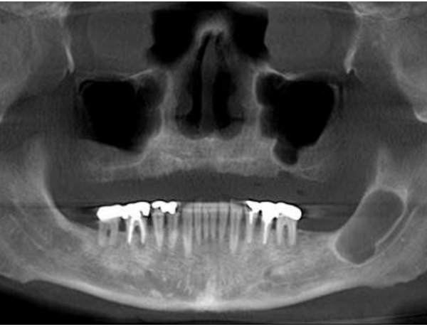
Radiological examination
• Maxilla. Suitable views are:
• Periapicals and oblique occlusals
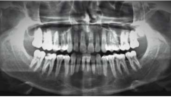
• Panoramic radiograph or lateral oblique
• Occipitomental (OM)
• True lateral (anterior maxilla).
• Mandible. Suitable views are:
• Periapicals and true occlusals
• Panoramic radiograph or lateral oblique
• Posteroanterior (PA) of mandible.
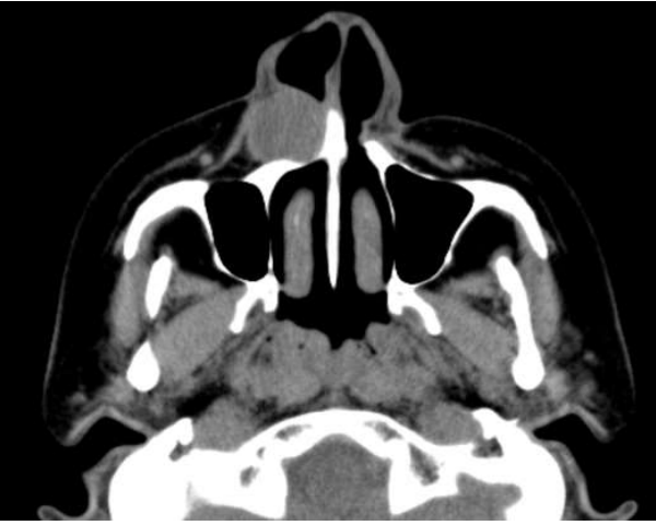
• Computed tomography (CT) may be useful in
• Planning surgery of large cysts, particularly in the
• Posterior maxilla.
• Radiological signs
• Classically, cysts appear as well-defined round or
• Ovoid radiolucencies, surrounded by a well-defined
• Margin.
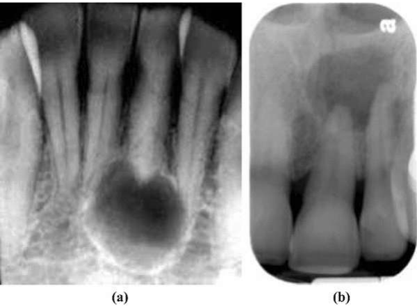
Radicular cyst
A well-defined, round or ovoid radiolucency is associated with the root apex
Residual cyst
• The residual cyst has a well-defined, round/ovoid radiolucency in an edentulous area
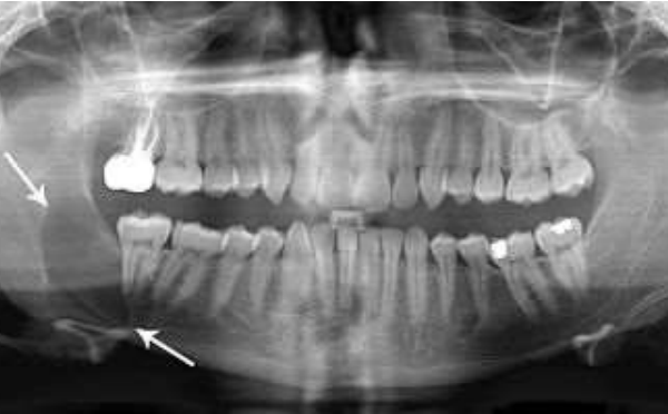
Odontogenic keratocyst
• 4 – 12% of all odontogenic cysts (often compared to odontogenic cysts even though WHO classifies as tumor)
• Peaks in second and third decade of life, but can occur over wide age range
• 90% are solitary
• Multiple tumors seen in Nevoid Basal Cell Carcinoma Syndrome / Gorlin Syndrome •Mandible most commonly involved (65 – 85% of KCOT)
• Most common site: posterior mandible
• Not uncommonly, but not exclusively associated with impacted teeth
• Rarely occurs in soft tissue
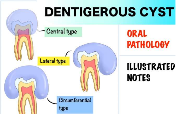
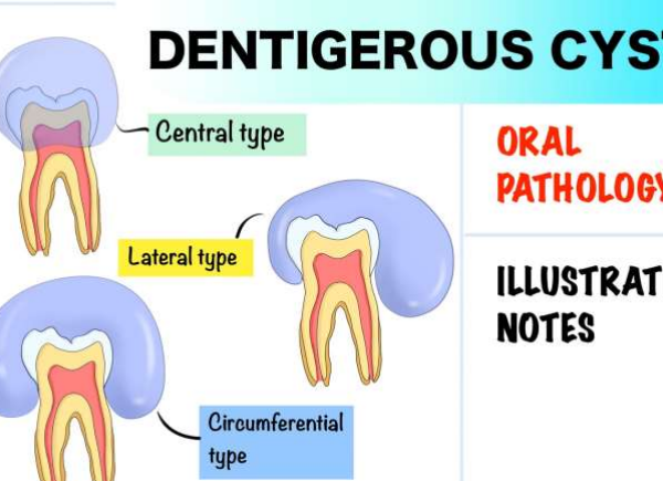
Dentigerous cyst
• In dentigerous cysts, there is a pericoronal radiolucency greater than 3– 4 mm in width that is suggestive of cyst follicle
Eruption cyst
• The extra-bony position of the eruption cyst means
• that the only radiological sign is likely to be a soft
• tissue mass.
Gingival cysts
• Gingival cysts are commonly found in neonates but are rarely encountered after 3 months of age • Gingival cysts are lined by stratified squamous parakeratotic epithelium. In neonates and infants, the cysts are typically between 2 and 5mm in diameter
Nasopalatine cyst
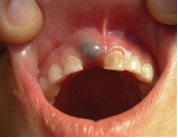
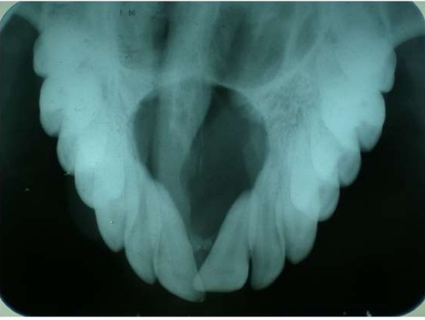
• The nasopalatine cyst appears as a well-defined round radiolucency in the midline of the anterior maxilla
Nasolabial cyst
• As the nasolabial cyst is a soft tissue lesion, radiography may reveal nothing
Solitary bone cyst
• The solitary bone cyst appears as a well-defined but non-corticated radiolucency.
• Typically, it has
• little effect on adjacent structures and ‘arches’ up
• between the roots of teeth
Aneurysmal bone cyst
• The aneurysmal bone cyst typically presents as a fairly well-defined radiolucency
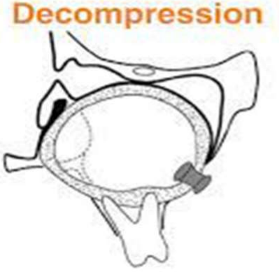
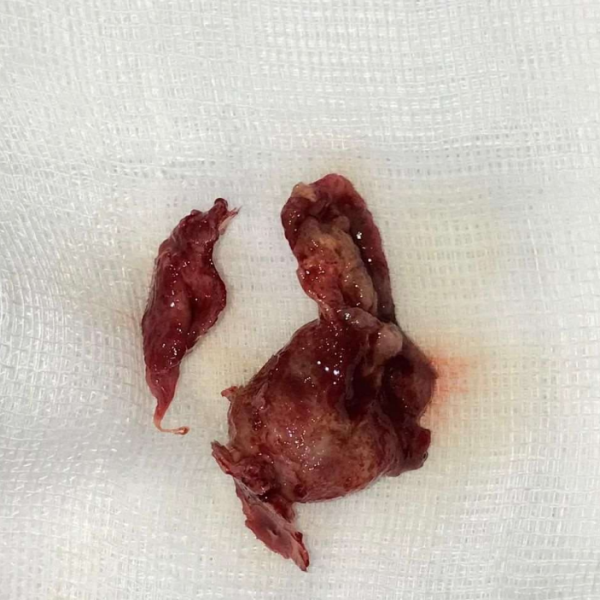
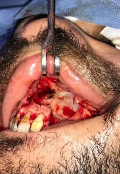
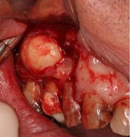
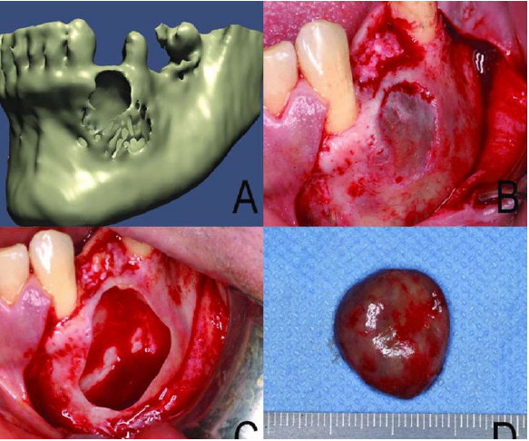
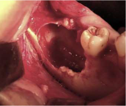
Surgical management of cysts
• Surgical management of cysts generally implies enucleation, but occasionally marsupialization is the technique of choice
Enucleation
• involves the removal of the whole cyst, including the epithelial and capsular layers from the bony walls of the cavity
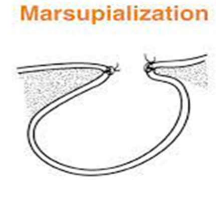
Marsupialisation
• Marsupialisation is a simple operation that may be performed under local anaesthesia in which a window is cut and removed from the cyst lining